2025: Volume 6, Issue 1
Past Issues
 Abstract
  PDF
PDFSirenomelia (Mermaid Malformation): A Case Report and Review of the Literature
Hemza Guellouh1,2,*, Fadila Bendaoud1,2
1Department of Pediatrics and Neonatology, Specialized Mother and Child Hospital, Batna, Algeria
2Faculty of Medicine, Batna 2 University, Algeria
*Corresponding author: Hemza Guellouh, Department of Pediatrics and Neonatology, Specialized Mother and Child Hospital, Batna, Algeria & Faculty of Medicine, Batna 2 University, Algeria, Phone: +213 550 97 94 10, E-mails: [email protected]; [email protected]
Received Date: February 07, 2025
Published Date: February 20, 2025
Citation: Guellouh H, et al. (2025). Sirenomelia (Mermaid Malformation): A Case Report and Review of the Literature. Neonatal. 6(1):23.
Copyrights: Guellouh H, et al. ©?(2025).
ABSTRACT
Sirenomelia is a rare congenital malformation, characterized by early fusion (symelia) of the lower limbs, either complete or incomplete, resulting in a single limb resembling a mermaid. It is frequently associated with other malformations such as bilateral renal agenesis, anal imperforation, lumbosacral agenesis, single umbilical artery, renal or external genital dysplasia, as well as cardiac or neurological abnormalities. This malformation is fatal due to pulmonary hypoplasia or complications from associated anomalies. In this case, we report a rare case of sirenomelia in a 1-day-old newborn, hospitalized for the management of sirenomelia diagnosed postnatally, who presented with total symelia, absence of anus and genital organs, agenesis of internal organs, both kidneys, and the renal tract, and passed away on the second day of life, to highlight a rare malformation and the importance of prenatal diagnosis for obstetric monitoring, as well as the need to raise awareness about genetic counseling.
Keywords: Sirenomelia, Mermaid Syndrome, Congenital Malformation, Malformation Rare, Neonatology
INTRODUCTION
Sirenomelia is a rare congenital malformation [1], characterized by early fusion (symelia) of the lower limbs, either complete or incomplete, resulting in a single limb resembling a mermaid.
It is frequently associated with other malformations such as bilateral renal agenesis, anal imperforation, lumbosacral agenesis, single umbilical artery, renal or external genital dysplasia, as well as cardiac or neurological abnormalities [1-3].
This malformation is fatal due to pulmonary hypoplasia or complications from associated anomalies [2].
In this case, we report a rare case of sirenomelia in a 1-day-old newborn, hospitalized for the management of sirenomelia diagnosed postnatally, to highlight a rare malformation and the importance of prenatal diagnosis for obstetric monitoring, as well as the need to raise awareness about genetic counseling.
CASE REPORT
The newborn Z, of indeterminate sex, aged 1 day, was born from a 38-week pregnancy via cesarean section. birth weight: 1700 g, Apgar scores: 7/10, 8/10. Mother's history: G1/P0, 37-year-old primiparous woman.
Clinical Examination at Birth: The newborn presented with: good skin and mucosal coloration, Eupneic respiration, good axial and segmental tone, normotensive anterior fontanelle, normal cardiac and pulmonary auscultation, soft abdomen, patent choanae and esophagus, total fusion of both lower limbs (symelia), absence of anus and external genitalia, positive sucking reflex.
Paraclinical Examinations: Skeletal X-ray: Showed the presence of bones of both lower limbs without associated skeletal anomalies. Abdominopelvic ultrasound: Revealed agenesis of internal organs, bilateral renal agenesis, and renal tract agenesis. Echocardiography: No abnormalities detected.
Management and Outcome: The newborn received basic intravenous fluid therapy with 10% glucose solution supplemented with electrolytes (calcium gluconate, NaCl, KCl). One day after birth, the newborn developed moderate respiratory distress, which progressively worsened, leading to severe respiratory distress. Two days after birth, the newborn passed away due to severe respiratory distress (Figures 1,2&3).
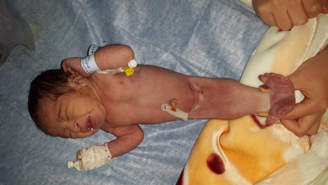
Figure 1. Front view of a newborn with sirenomelia.
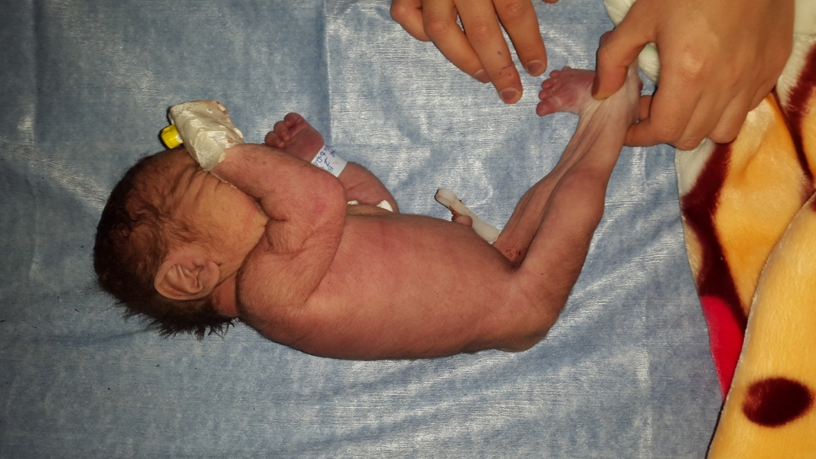
Figure 2. Side view of a newborn with sirenomelia.
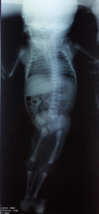
Figure 3. Frontal X-ray of the newborn's skeleton showing the presence of 2 femurs, 2 tibias, and 2 fibulas (Type 1 according to Stocker and Heifetz).
DISCUSSION
Sirenomelia, also known as mermaid syndrome, mermaid malformation, or sirenomelia sequence [2], is a rare malformation with an incidence of 1/600,000–1,000,000 [3]. It was first described in 1945 [3]. The risk is lower in twin pregnancies but significantly higher in monozygotic twins, with a risk up to 100 times greater compared to singleton or dizygotic twin pregnancies [2].
In our case, the newborn was from a singleton pregnancy. It is important to consider this malformation sequence during ultrasound follow-up, particularly in twin pregnancies. The malformation may be linked to an unknown genetic anomaly or other contributing factors [2].
Sirenomelia can be diagnosed intrauterinely from 22 weeks of gestation [1]. Prenatal color Doppler imaging is valuable as it can aid in diagnosis, especially in cases of bilateral renal agenesis [1]. In our case, the diagnosis was made postnatally following a clinical examination of the newborn. A notable factor for the mother was that she came from a rural area with no specialized gynecologist. Her pregnancy was monitored by a general practitioner trained in obstetric ultrasound. This highlights the importance of accessible, high-quality prenatal follow-up by well-trained and informed physicians, as delayed or missed diagnoses can significantly impact prognosis. Additionally, raising awareness among expectant mothers about the necessity of continuous and quality follow-up is crucial.
In cases of early detection of sirenomelia, pregnancy termination may be an option. On a global scale, pregnancy termination occurs in 49.5% of cases. This decision depends on two key factors: early detection of the malformation and parental consent. In our case, there was no prenatal diagnosis, making termination impossible. Furthermore, cultural and religious beliefs in the local context prohibit such practices, making parental consent difficult even in cases of early diagnosis.
This anomaly results in a malformation sequence, including complete or incomplete fusion of the lower limbs (symelia); gastrointestinal malformations such as anal imperforation, anal atresia, omphalocele, esophageal atresia, and a blind-ending colon; a single umbilical artery; renal malformations such as renal dysplasia, bilateral renal agenesis, oligohydramnios, or polyhydramnios; skeletal malformations, dysplasia of external genital organs, lumbosacral agenesis, cardiac malformations, and neurological abnormalities [1-3].
In our newborn, clinical examination revealed complete fusion of the lower limbs (symelia), with the presence of both lower limb bones on skeletal radiography. External genital organs were absent, and the infant had anal imperforation, but no other clinically detectable anomalies. Severe gastrointestinal malformations were noted, though not all those described in the literature were present. Due to their frequency, such malformations should be systematically investigated. The umbilical artery appeared normal without anomalies. Given the strong association between symelia and renal malformations, we performed an abdominopelvic ultrasound, which revealed bilateral renal agenesis, renal tract agenesis, and even agenesis of internal abdominal organs. However, echocardiography showed no cardiac malformations, and neurological imaging as well as chest radiography were unremarkable. These findings emphasize the necessity of comprehensive radiological exploration when symelia is clinically observed, particularly to assess potential renal, gastrointestinal, skeletal, cardiac, and neurological malformations.
Regarding the classification of sirenomelia, based on the presence or absence of lower limb bones, Stocker and Heifetz proposed a classification system with seven types [1] (Table 1). Our newborn had type 1 sirenomelia, as both femurs, tibias, and fibulas were visible on skeletal radiography.
Other rare genetic syndromes may occasionally include symelia, such as the Selign-Bennacerraf-Green syndrome and Rudd-Klimek syndrome. It is also essential to differentiate sirenomelia from caudal regression syndrome, which is typically associated with maternal diabetes and is characterized by sacral and lumbar vertebral agenesis along with neurogenic atrophy of the lower limbs. Some authors even consider mermaid syndrome to be a severe form of caudal regression syndrome [4].
Regarding prognosis, more than 50% of newborns with sirenomelia die in utero, and only 1% survive beyond one week of life. Most of these newborns succumb within the first year due to complications, primarily pulmonary or renal in nature. In our case, the newborn developed moderate respiratory distress on the first day of life, which progressively worsened and became severe, leading to death on day 2. Due to the severity of these malformations, sirenomelia is incompatible with life, making death inevitable. In such situations, it is crucial to thoroughly explain the malformation sequence to parents, its severity, potential complications, and its incompatibility with life. This ensures psychological preparedness and encourages parental adherence to follow-up care.
It is also important to note that in certain countries, particularly in Africa, due to lack of awareness and mystical or religious beliefs, mothers of affected infants are often accused of being cursed or practicing witchcraft [5]. This highlights the need to educate families about the probable genetic origin of this malformation [2] to prevent unnecessary and unjustified psychological distress, especially in families with limited education. Additionally, parents should be informed about the importance of genetic counseling to assess future obstetric risks.
Table 1. Classification of sirenomelia according to Stocker and Heifetz [1]
|
Type |
Description |
|
1 |
Presence of 2 femurs, 2 tibias, 2 fibulas |
|
2 |
Presence of a single fibula |
|
3 |
Absence of fibula |
|
4 |
Partially fused femurs with fused fibulas |
|
5 |
Partial fusion of fibulas |
|
6 |
Presence of a single femur with a single tibia |
|
7 |
Presence of a single femur with absence of tibia |
CONCLUSIONS
Sirenomelia is a rare malformation that can be diagnosed early prenatally through proper obstetric monitoring of pregnancy. It is fatal due to associated malformative complications. When diagnosed early in utero, pregnancy termination may be an option. Given this condition, genetic counseling for the parents is recommended.
AUTHORSHIP CRITERIA
- Hemza Guellouh: Concept and design of study or acquisition of data or analysis and interpretation of data;
- Hemza Guellouh: Drafting the article or revising it critically for important intellectual content; and
- Hemza Guellouh, Fadila Bendaoud: Final approval of the version to be published. Authors should provide a description of contributions made by each of them towards the manuscript.
Guarantor Author: Hemza Guellouh.
ACKNOWLEDGEMENTS
No Acknowledgements.
CONFLICTS OF INTEREST/COMPETING INTERESTS
Conflicts of interest/ Competing internets: None related to this article.
GRANT INFORMATION
The author(s) received no specific funding for this work.
REFERENCES
- Ladure H, D’Hervé D, Loget P, Poulain P. (2006). Diagnostic anténatal d’une sirénomélie. J Gynecol Obstet Biol Reprod (Paris). 35(2):181-185.
- Zeki Ya?ar, M, Yusuf AA, Hassan FM, Ali AA, Roble MA. (2022). Mermaid syndrome: A case report in Somalia. Annals of Medicine and Surgery. 76:103533.
- At?c? A, Çelikkaya ME, Arslan S, El Ç, Akçora B. (2018). Sirenomelia/mermaid syndrome without imperforate anus in a premature infant. Journal of Pediatric Surgery Case Reports. 30:46-47.
- Samal SK, Rathod S. (2015). Sirenomelia: The mermaid syndrome: Report of two cases. J Nat Sci Biol Med. 6(1):264-266.
- Lubala TK, Mukuku O, Mutombo AM. (2014). Sirénomélie (Mermaid Syndrome): description du premier cas congolais et revue de la littérature. Pan Afr Med J. 17:162.