2023: Volume 4, Issue 1
Past Issues
 Abstract
  PDF
PDFComplex Body Wall Anomalies in a Survived Patient: A Case Report
Ahmed Elkordy1,*, Anne Marie Tuyisenge2, Maha Saleh3, Anita Cheng4
1Clinical Fellow, Department of Pediatrics, Division of Neonatal-Perinatal Medicine, Western University, Canada
2Department of Pediatrics, Division of Neonatal-Perinatal Medicine, Western University, Canada
3Assistant Professor, Department of Pediatrics, Division of Genetics, Western University, Canada
4Assistant Professor, Department of Pediatrics, Western University, Canada
*Corresponding Author: Ahmed Elkordy, Clinical Fellow, Department of Pediatrics, Division of Neonatal-Perinatal Medicine, Western University, Canada; Email: [email protected]
Received Date: January 5, 2023
Publication Date: May 8, 2023
Citation: Elkordy A, et al. (2023). Complex Body Wall Anomalies in a Survived Patient: A Case Report. Neonatal. 4(1):9.
Copyright: Elkordy A, et al. © (2023).
ABSTRACT
Complex body wall anomalies (CBWA) is a congenital anomaly that carries a poor prognosis and can present diagnostic challenges due to the overlapping nature of its related terms – including Limb body wall complex (LBWC) and Body stalk anomaly (BSA). It is essential to achieve an accurate diagnosis of these anomalies, as variations of the diagnosis can have significant implications about the baby's prognosis. The CBWA classification system has been developed to differentiate categories based on anatomy, which is helpful in reaching more accurate diagnoses. In this case report, the baby's condition can be categorized according to Martin-Alguacil’s CBWA classification as BSA VII or LBWC II [1]. BSA with abdominoschisis (Type III and Type VII) were considered LBWC Type II as described by Martín?Alguacil, et al. [2]. We describe a case report of a surviving infant with CBWA who is currently 37 months old and survived without requiring ongoing life-sustaining interventions. This case highlights that a wide range of prognoses for this condition is possible and should therefore be considered when giving antenatal recommendations.
Keywords: Complex body wall anomalies; congenital defect; APGAR scores; NICU
INTRODUCTION
CBWA is a rare, sporadic, congenital defect described by a combination of at least two of three characteristics: limb defects, anterior body wall defects, and exencephaly or encephalocele, with or without facial clefts [3]. Different hypotheses proposed to explain the pathogenesis of limb body wall complex include early amnion disruptions, embryonic dysplasia, and vascular disruption in early pregnancy [3]. The prevalence reported ranges from 0.4 to 3.2 per 100,000 live births and stillbirths [4]. However, the true prevalence may be underreported if not accounting for spontaneous fetal losses and termination of pregnancies.
Prenatal diagnosis of CBWA is made possible with routine antenatal anatomy ultrasound screening. Accurate antenatal diagnosis is important, as the prognosis is different according to the constellation of findings detected. We present an interesting case that highlights a wider spectrum of CBWA, where survival is possible. This element of uncertainty is important to consider when counseling parents antenatally and illustrates the importance of multidisciplinary care to provide perinatal decision making and management.
CASE REPORT
The mother was 22 years old, G2A1, seroprotective. She had been referred to the multidisciplinary prenatal clinic due to an anatomy scan at 18 weeks showing fetal omphalocele. She had declined routine 1st-trimester screening. Repeated ultrasound at 20+3 weeks showed a large omphalocele with complex cystic areas containing the liver. The ultrasound also showed scoliosis, absent left leg with the right leg seen with an abnormal orientation of the foot. Fetal echo showed a qualitatively small ascending aorta and the possibility of significant left-sided pathology. There was a nuchal fold of 6.6 mm, and the baby was small for gestational age (estimated fetal weight <3rd percentile). The remainder of the anatomy was normal.
The parents were counseled by the maternal-fetal medicine, neonatal-perinatal medicine, and genetics teams that the findings likely represented CBWA, which carries a poor prognosis and is usually lethal. The differential diagnoses included trisomy 13 or trisomy 18. Parents were offered further genetic testing, which they declined. The option of termination of pregnancy was also declined. A repeat ultrasound at 31 weeks confirmed these findings but with no appearance of scoliosis. The parents decided to proceed with the pregnancy and for full life-sustaining therapies for the baby until they were able to visually confirm ultrasound findings postnatally. They had agreed that if the baby was truly found to have only one leg, they would opt for comfort care only.
A male infant was born via elective cesarean section at 38 weeks and 3 days gestation. Birth weight was 2630g (11th percentile); the length was 45 cm (3rd percentile), and head circumference was 35 cm (64th percentile). He cried after birth but had some respiratory distress with work of breathing and was placed on CPAP. Parents were brought into the room to see the baby. Despite that he was confirmed to only have one leg, upon seeing him relatively stable in that he was making good respiratory efforts, they decided to continue full life-sustaining therapy. Due to ongoing respiratory distress and work of breathing despite CPAP, the baby was intubated and ventilated.
Physical examination revealed neither facial dysmorphism nor orofacial clefting. Upper limb examination was normal. He had a normal appearance of the chest and a large omphalocele. Lower limb examination showed an absent left leg at the level of the hip and an externally rotated right leg with right club foot. There was no scoliosis. He had a 2 vessel umbilical cord distal to the large omphalocele. Unfortunately, we were unable to get a histological study on the umbilical cord, which can be important in determining the diagnosis. There was no thoracoschisis. The remainder of his physical exam was unremarkable. These findings are in keeping with a diagnosis of LBWC type II [1].
In the NICU, he was neurologically appropriate and his head ultrasound and brain MRI were normal. He was successfully extubated on day 6 and weaned to room air at 2 weeks. The pediatric surgery team planned for staged closure of the omphalocele, using sulfasalazine topically. He was able to bottle feed with appropriate weight gain. An echocardiogram showed a small PDA and bicuspid aortic valve. He was discharged home after 24 days.
At 4 months of age, the omphalocele was completely reduced by following frequent dressing and wrapping. He continued to gain weight appropriately. There was a plan was to proceed with surgical repair of the abdominal wall at 2 years of age. He had intermittent constipation. At 5 months of age, he was diagnosed with bilateral cataracts, which were surgically treated. A congenital cataract genetic panel, as well as a chromosomal microarray, were ordered and revealed no pathogenic copy number variations. At 13 months of age, he showed mild motor delay due to the absent leg, but had normal language and social development. He was tolerating oral feeding, stooling, and voiding with no concerns. Overall, at 35 months of age, the baby is thriving and described by parents to have a good quality of life.
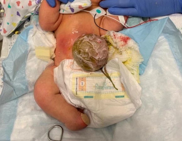
Figure 1: At birth. Omphalocele dressed, left leg absent, right leg abnormally rotated.
.jpg)
Figure 2: 2 months. Sulfasalazine cream applied to omphalocele.
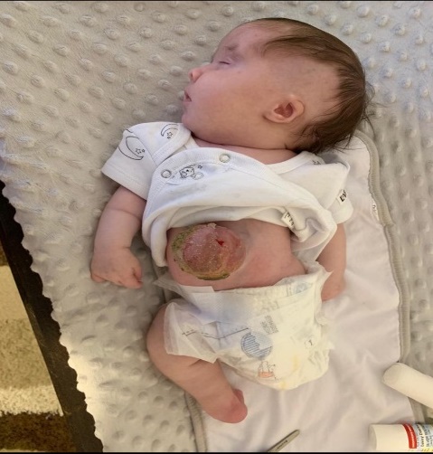
Figure 3: 3 months. Sulfasalazine cream continued to be applied to omphalocele.
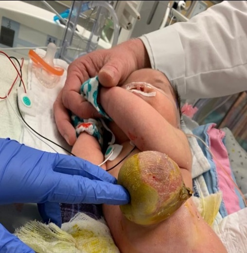
Figure 4: 4 months. Omphalocele treated with topical Flamazine and wrapped with Kling dressing.
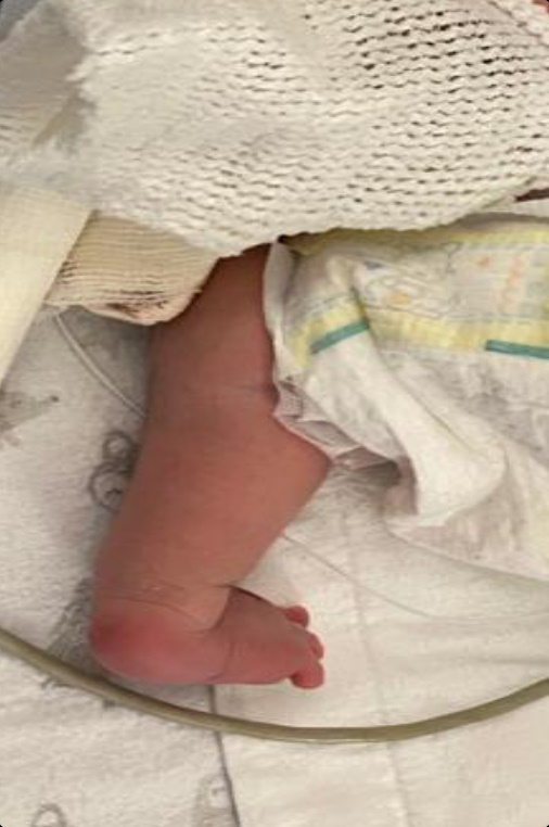
Figure 5: 18 months. 1 week post omphalocele reduction surgery, covering the omphalocele with dry gauze. Right leg shown externally rotated.
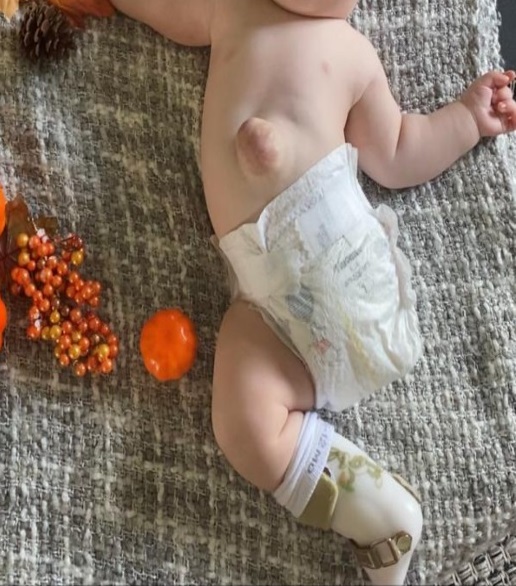
Figure 6: 19-months. Old omphalocele site continuing to heal; right leg in cast to mitigate external rotation.
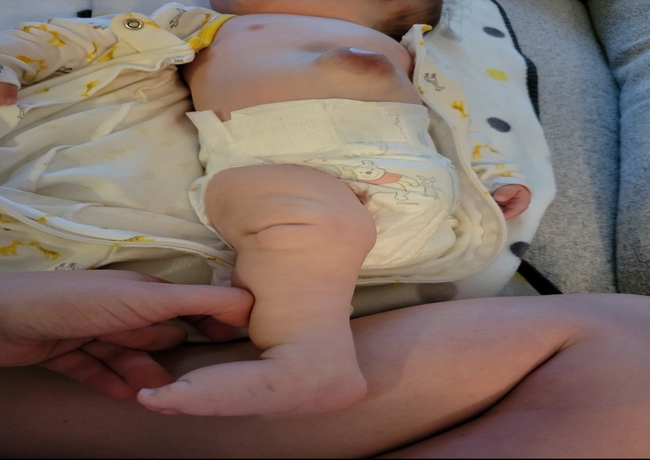
Figure 7: 2 years. Improvement in right leg external rotation. Old omphalocele site well healed.
DISCUSSION
Complex body wall anomalies (CBWA) is a rare congenital disease. There are some theories, like Tropin's amniotic band theory and Van Allen's vascular theory, which could explain the mechanism for the constellation of anomalies [5-8]. On the other hand, there is no clear evidence of genetic or teratogenic predispositions as causal factors [9].
Because of the wide spectrum of the features involved and variable prognoses, we support Martin-Alguaci, et al. [1] that classifying the cases with a more specific approach is crucial in providing more accurate prognostic information to parents and healthcare providers. It can help in managing parental expectations and counseling them about the possible outcomes.
In the past, CBWA was often considered a lethal condition and pregnancy termination was the plan for many cases [6]. There are several reported cases of non-surviving fetuses described by Martín-Alguacil, et al. [1], including babies with abdominoschisis, spinal defects, anomalous umbilical cord, and structural limb defects. These cases include those reported by Chen et al. (2007) [10], Durga & Renukadevi (2016) [11], Gulczy?ski, et al. (2019) [12] and Gupta, et al. (2015) [13].
Our case is the oldest surviving published case of CBWA to date. There are only a few other reported cases of infants surviving the neonatal period with CBWA, including those described by Lazaroni, et al. (2016) [14], Kundal, et al. (2015) [15], and Kanamori, et al. (2007) [16]. Like our case, these surviving patients also had normal face, brain, and heart morphology, and did not exhibit pulmonary hypoplasia or a short umbilical cord. However, there are key differences between our case and these others. For instance, the case described by Lazaroni, et al. had a bulky ruptured omphalocele (liver and gall bladder, intestines and pancreas, and externalized testis), pelvic asymmetry, and atrophy of the lower limbs with clubfoot, and only reported 3-month survival. Kundal, et al.'s case had a major omphalocele and an absent whole left lower limb with an abnormally fused right foot. Unfortunately, there was no follow-up information available for this case. In addition, we observed a unique finding of cataract in our case, which has not been reported in other similar cases of CBWA. To investigate the genetic basis of this finding, we offered a congenital cataract next generation sequencing panel that included 113 genes. However, the results of this panel were negative, suggesting that further investigation may be needed to better understand the genetic underpinnings of this unique finding in our case. Our case represents a rare and important example of long-term survival in CBWA, and our findings suggest that certain factors, such as normal face, brain, and heart morphology, can play an important role in determining outcomes in these cases.
The antenatal diagnosis of CBWA (including LBWC) is possible, detecting the presence of abdominal wall defects, kyphoscoliosis, and limb abnormalities during the ultrasound examination in the first trimester. Additionally, increased maternal serum alpha-fetoprotein can be an early clue in the diagnosis of CBWA, while karyotyping may not add a relevant value as it is usually normal [6]. It has been a general agreement in many articles that CBWA is considered a lethal anomaly, but reconsidering it as a congenital anomaly with a usually poor prognosis would be more accurate to reflect the spectrum of disease [17]. In many cases, clinicians preferred early diagnosis followed by offering pregnancy termination as a standard treatment for CBWA [6,18]. However, in our case, parents chose to continue the pregnancy and to defer the decision regarding the goals of care postnatally. After the delivery, the decision was confirmed by the parents to continue resuscitation and provide full life sustaining therapy for the baby.
The ethical aspects of this case are important; when medical staff is confronted with the challenge of treating children with rare and complex diseases, especially those that include narrow or often poor outcomes, as is considered in CBWA. The parents’ decision was respected, despite the perception that prolonged hospitalization of the baby may cause both an emotional and financial burden on the family and the healthcare system. Multidisciplinary care was fundamental, both in supporting parents through decision making processes and collaborating to meet the many medical needs of this baby. The course of our case was unexpectedly favorable, likely because there were no thoracic or brain abnormalities [19-23].
CONCLUSION
Our case highlights that not all variants of CBWA are lethal, especially in the absence of thoracic and brain involvement. We report a baby who has survived 37 months and is currently thriving. This case demonstrates the importance of prenatal diagnoses and multidisciplinary perinatal care for accurate counseling, considering a spectrum of outcomes. It is also important to consider the ethical challenges in shared decision-making with parents regarding goals of care.
REFERENCES
Martín-Alguacil N. (2020). Anatomy-based diagnostic criteria for complex body wall anomalies (CBWA). Mol Genet Genomic Med. 8(10):e1465.
Martín?Alguacil N, Avedillo L. (2020a). Body stalk anomalies in pig-Definition and classification. Mol Gen Gen Med. 13:e1227.
Kocherla K, Kumari V, Kocherla PR. (2015). Prenatal diagnosis of body stalk complex: A rare entity and review of literature. Indian J Radiol Imaging. 25(1):67–70.
Bugge M. (2012). Body stalk anomaly in Denmark during 20 years (1970–1989). Am J Med Genet A. 158A:1702–1708.
Van Allen MI, Curry C, Gallagher L. (1987). Limb body wall complex: I. Pathogenesis. Am J Med Genet. 28:529–548.
Chikkannaiah P, Dhumale H, Kangle R, Shekar R. (2013). Limb body wall complex: a rare anomaly. J Lab Phy. 5 (1):65–67.
Torpin R. (1965). Amniochorionic mesoblastic fibrous strings and amnionic bands: Associated constricting fetal malformations or fetal death. Am J Obstet Gynecol. 91:65–75.
Pumberger W, Schaller A, Bernaschek G. (2001). Limb-body wall complex, a compound anomaly pattern in body-wall defects. Pediatr Surg Int. 17:486-490.
Chen CP, Lin CJ, Chang TY, Hsu CY, Tzen CY, Wang W. (2007). Second?trimester diagnosis of limb?body wall complex with literature review of pathogenesis. Genetic Counseling. 18:105–112.
Durga, R., & Renukadevi, T. K. (2016). Amniotic Band Syndrome-A dreaded condition. J Clin Diagn Res. 10:QD04–QD05.
Gulczy?ski J, ?wi?tkowska-Freund M, Paluchowski P, Hermann-Okoniewska B, I?ycka-?wieszewska E. (2019). Limb body wall complex–The history of the entity and presentation of our series of cases. Polish J Pathol. 70:33–41.
Gupta K, Venkatesan B, Chandra T, Rajeswari K, Devi TK. (2015). Amniotic band syndrome with sacral agenesis and umbilical cord entrapment: A case report emphasizing the value of evaluation of umbilical cord. J Radiol Case Rep. 9:12–19.
Lazaroni T, Cruzeiro PCF, Piçarro C, Victoria AM, Filho FMB, Tatsuo ES, et al. (2016). Body stalk anomaly: Three months of survival. Case report and literature review. J Pediatr Surg Case Rep. 14:22-25.
Kundal V, Gajdhar M, Kundal R, Ahmed R, Agrawa L. (2015). Omphalocele Major with Absent Lower Limb. Indian J Surg. 77(1):70-71.
Kanamori Y, Hashizume K, Sugiyama M, Tomonaga T, Takayasu H, Ishimaru T, et al. (2007). Long-Term Survival of a Baby with Body Stalk Anomaly: Report of a Case. Surg Today. 37:30–33.
Pardo Vargas RA, Aracena M, Aravena T, Cares C, Cortés F, Faundes V, et al. (2016). Congenital anomalies of poor prognosis. Genetics Consensus Committee. Rev Chil Pediatr. 87(5):422-431.
Van Allen MI, Curry C, Walden CE, Gallagher L, Patten RM. (1987). Limb body wall complex: II. Limb and spine defects. Am J Med Genet. 28:549-556.
Levy R, Lacombe D, Rougier Y, Camus E. (2007). Limb body wall complex and amniotic band sequence in sibs. Am J Med Genet. 143A:2682-2687.
Hartwig NG, Vermeij-Keers C, De Vries HE, Kagie M, Kragt H. (1989). Limb body wall malformation complex: An embryologic etiology? Hum Pathol. 20:1071–1077.
Luehr B, Lipsett J, Quinlivan JA. (2002). Limb-body wall complex. A case series. J Matern Fetal Neonatal Med. 12:132-137.
Russo R, D’Armiento M, Angrisani P, Vecchione R. (1993). Limb body wall complex a critical review and a nosological proposal. Am J Med Genet. 47(06):893–900.
Adeleke O, Gill F, Krishnan R.. (2022). Rare Presentation of Limb–Body Wall Complex in a Neonate. Case Report and Review of Literature. AJP Rep. 12:e108–e112.
Borade A, Prabhu AS, Prabhu GS, Prabhu SR. (2009). Limb Body Wall Complex (LBWC). Pediatric Oncall J. 6:45-46.